Tag Archives: imaging

Medis: The new 4D Flow Module reduces the complexity of HeartMRI scans substantially

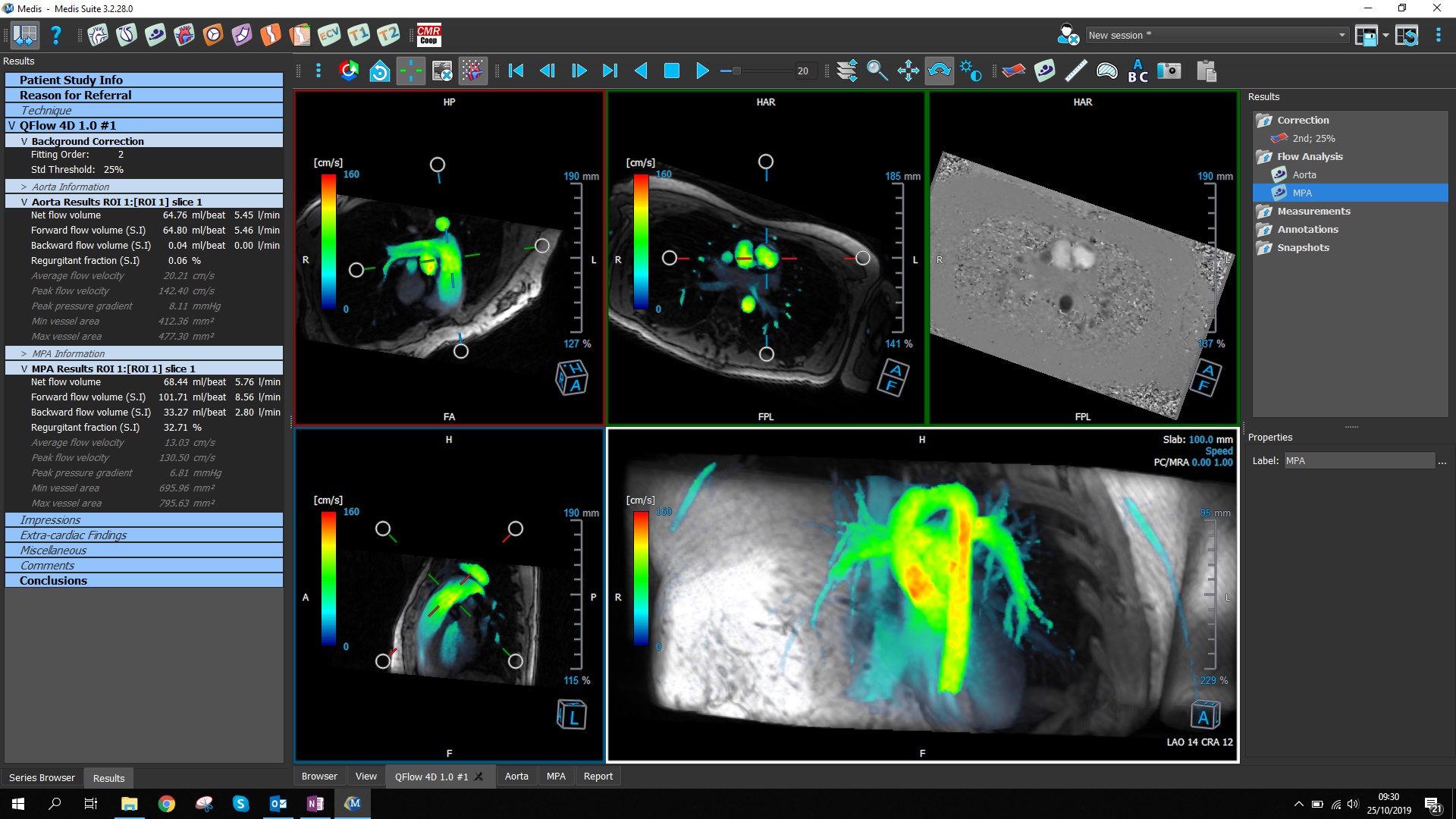
LEIDEN, Netherlands, 4-Nov-2019 — /EPR HEALTHCARE NEWS/ — Innovative cardiovascular imaging technology solutions developer Medis today announced a new module for the company’s Medis Suite MR. The new 4D Flow module is intuitive and easy to use, and provides essential, practical tools to enable 4D flow to be incorporated into clinical routine and to allow the measurement of flow volumes from a single 4D flow scan instead of multiple 2D flow scans – reducing complexity and easing the process.

“With 2D flow scans, reconstructions must be planned while the patient is on the table,” said Hans Brons, CEO. “For complex scan protocols, this can be tedious and challenging – and the post-processing can be both time-consuming and complex. With the help of this new module for Medis Suite MR, it is now possible to acquire flow in 4D from a single scan and to create flow plane reconstructions retrospectively, allowing the complexity of HeartMRI scans to be reduced substantially – and also reducing the time it takes. This is especially relevant for paediatric HeartMRI units as well as units scanning patients with grown up congenital heart disease.”

Among its numerous differentiators, the new module is advantaged by its ease of use with an intuitive GUI, as well as single click noise removal and single click background offset correction.

As well as reducing the time taken, the new 4D Flow module supports the experienced MR technician in being more effective, more productive and able to deliver better diagnostic information. In addition to reducing the time taken, the new module is straightforward to learn and use for measuring flows in a variety of vessels, allowing the efficient incorporation of 4D Flow post processing in daily clinical practice.

The new 4D Flow module for Medis Suite MR is part of a wide-ranging suite of enhancements for the platform, including the 3D View module – further reducing the time needed for post-processing. In the 3D View module, a double caliper has been added for simple measurements. In addition, loading speed has been increased and it is now also possible to create straightened CPRs (curved planar reformats). Finally, Medis Suite AutoQ pre-processing can now be enabled to run autonomously, initiating Deep Learning contour detection and other advanced algorithms automatically.

Based on over 15 years of experience with cardiac MR, and over 30 years of cardiovascular medical image analysis in general, Medis Suite MR is a vendor-independent post-processing solution for HeartMRI cases. The workflow in Medis Suite MR includes a practical HeartMRI viewer, advanced clinical applications and convenient reporting all in one, making it highly efficient to work with. Seamlessly embedded in the workflow are advanced clinical applications considered best-in-class by many, such as the renowned QMass® and QFlow® applications. In addition, highly innovative and cutting edge research apps such as QStrain RE and QMap RE are also integrated.

Able to run on any workstation, Medis Suite MR is simple to integrate within the hospital IT environment. Connecting it to the DICOM network is straightforward.

4DFlow is cleared for market in the US Market. Clearance approvals for Australia, Brazil, Canada and Europe are pending.

Further information on Medis and its products is available at https://www.medis.nl/

SOURCE: EuropaWire

Multispectral Optoacoustic Tomography (MSOT) can now more reliably detect early stages of inflammation

GERMANY, Mar-30-2017 — /EPR HEALTHCARE NEWS/ — Ulcerative colitis and Crohn’s disease are the most common inflammatory bowel diseases (IBD). Globally, more than 4 million people suffer from recurring inflammation of the intestinal mucosa. To date, non-invasive methods have not been able to detect these forms of IBD reliably. Researchers at the University Hospital Erlangen (Germany) have now shown for the first time how Multispectral Optoacoustic Tomography (MSOT) can more reliably detect early stages of inflammation when compared with other non-invasive diagnostic methods. These results have recently been published in the New England Journal of Medicine.

Evaluating the inflammatory activity of the intestines early and accurately helps to prevent complications associated with IBD and guide an optimal therapy regime. Currently, doctors frequently rely on an invasive endoscopy procedure to assess the intestinal mucosa of patients with IBD. Additionally, non-invasive imaging methods are increasingly used in the clinic. For example, ultrasound allows clinicians to examine the structure, thickness and blood perfusion of the intestinal wall with changes in perfusion patterns indicating an onset of inflammation.

“With the MSOT procedure, we additionally use laser light, by which we can detect inflammation earlier than previously possible with ultrasound”, explains Prof. Dr. Maximilian Waldner, Professor at the University Hospital Erlangen. “The earlier we detect signs of an upcoming flare – i.e., a change in blood content – the quicker we can react and start a preventive treatment”. The MSOT technology was developed by iThera Medical in Munich, and was first used on IBD patients last year.

The researchers of the University Hospital Erlangen have recently published their results in the renowned New England Journal of Medicine. “We examined 108 patients with Crohn’s disease using the MSOT technology and compared the results to established non-invasive diagnostic procedures as well as endoscopy and histology”, says Prof. Waldner. “We were able to demonstrate that hemoglobin values in the tissue measured with MSOT indicate very accurately the inflammatory activity in the intestines. The method appears to be superior to other non-invasive procedures, as MSOT for the first time was able to detect even low levels of inflammation without having to perform endoscopy”.

Dr. Ferdinand Knieling, a physician at the University Hospital Erlangen, adds: “We are hoping that this principle can be applied to many applications and, in particular, also be used to enable safe evaluations of children and adolescents. With this new examination method, many of the currently invasive procedures might become obsolete.”

In optoacoustic imaging, the physician scans the intestines transabdominally through the skin, applying pulsed laser light in the near-infrared region. The energy of the light absorbed in tissue is converted into acoustic signals which are then acquired by an ultrasound detector. Christian Wiest, CEO of iThera Medical, comments: “MSOT is a new diagnostic imaging modality that can detect changes in tissue composition associated with a variety of diseases, without the use of contrast agents. The technology has already been applied in other clinical feasibility studies, e.g. for the detection of melanoma metastasis in sentinel lymph nodes or for the assessment of suspicious breast lesions”.

The results of the IBD study at the University Hospital Erlangen were published on March 30th 2017 in the New England Journal of Medicine (Volume 376, Issue 13, pg.1292-1294).

SOURCE: EuropaWire